How Do You Know the Degree of a Dupuyten Contrature
Original Editor - Gwen Fritsche and Lauren Leimbach from Temple University's Bear witness Based Practice Project
Top Contributors - Lauren Leimbach, Gwen Fritsche, Admin, Lucinda hampton, Rachael Lowe, Kim Jackson, Chrysolite Jyothi Kommu, Laura Ritchie, Vidya Acharya, Scott A Burns, WikiSysop, Fasuba Ayobami, Lennert De Henau, Evan Thomas and Khloud Shreif
Introduction [edit | edit source]
Dupuytren contracture is a benign, myeloproliferative [1]progressive disease of the palmar fascia which results in shortening, thickening, and fibrosis of the fascia and aponeurosis of the palm.
- Dupuytren disease is predominantly a myofibroblastic disease that affects the mitt/fingers and results in contracture deformities.
- The most commonly affected digits are the third and fourth digits.
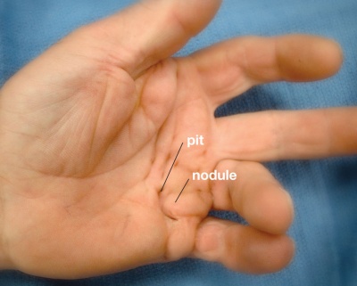
- The disease begins in the palm as painless nodules that form along longitudinal lines of tension.
- The nodules form cords that produce contracture deformities within fascial bands and tissues of the hand.
- Dupuytren contracture is ordinarily seen in whites and the disorder is often bilateral; when unilateral the correct side is more likely to be involved compared to the left.
- In many individuals, in that location is a family history with males being more likely to be affected than females.[ii] [3]
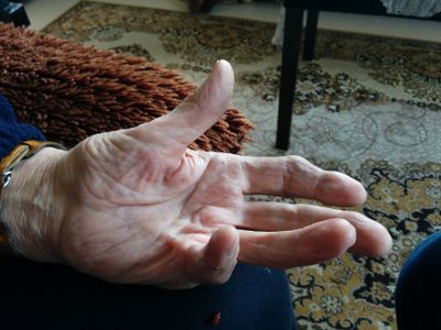
Paradigm ane: Clinical presentation of Dupuytren contracture[4]
Etiology [edit | edit source]
Dupuytren disease is a genetic disorder expressed in an autosomal dominant mode, just near frequently seen with a multifactorial etiology. Information technology is associated with diabetes, seizure disorders, smoking, alcoholism, HIV, and vascular disease[five].
Ectopic manifestations beyond the hand tin can be seen in Ledderhose affliction (plantar fascia), 10% to 30%; Peyronie disease (Dartos fascia of the penis), ii% to viii%; and Garrod disease (dorsal knuckle pads), 40% to l%.[5]
Epidemiology [edit | edit source]
This condition is common in populations of Northern European/Scandinavian descent. It is relatively uncommon in Southern European and South American populations and is rare in Africans and Asians. The disease affects men more severely than women. Males are affected past a 2:1 ratio compared to women. Younger age of onset is also associated with increased severity of illness progression. In Asian populations, the palm is more likely to be involved than the digits and thus often goes unnoticed.[five]
Pathological Procedure [edit | edit source]
The pathophysiology of Dupuytren affliction involves abnormal myofibroblastic growth in the hand.
- Blazon Three collagen predominates, which under a non-disease state would be Blazon I collagen.
- Dupuytren contracture progresses through three phases: (1) proliferative, (2) involution, and (3) residual. The proliferative phase has a characteristically high concentration of immature myofibroblasts and fibroblasts arranged in a whorled pattern. In the involution phase, fibroblasts get aligned in the longitudinal centrality of the hand following lines of tension. In the residual phase, relatively acellular collagen-rich chords remain causing contracture deformity.
- The disorder is not always progressive and in at least l-seventy% of patients, it may stabilize or even regress.
Several cords can develop which tin can crusade unique deformities of the hand.
- Pretendinous cords cause skin pitting and metacarpal phalangeal (MCP) articulation contracture.
- Natatory cords are responsible for webspace contractures.
- Spiral cords are the most important in the illness procedure and tin cause proximal interphalangeal (PIP) contracture.
Risk factors for increased severity and recurrence of disease after treatment include- male gender; onset earlier historic period 50; bilateral disease; sibling/parent interest; the presence of Garrod pads, Ledderhose, or Peyronies diseases.[two]
Clinical Presentation [edit | edit source]
Dupuytren contracture occurs slowly and typically progresses over the course of several years, simply tin can besides develop more rapidly over weeks or months.[6]
It typically affects older men of European descent. This condition almost commonly begins with thickening of the pare on the palm, resulting in a puckering or dimpled advent. As the status progresses, bands of fibrotic tissue form in the palmar area and may travel distally toward the fingers. This tightening and shortening eventually lead to the affected fingers being pulled into flexion. Dupuytren contracture typically occurs bilaterally, with i mitt being more than severely affected than the other.
Physical findings:
- Blanching of the skin when the finger is extended
- Proximal to the nodules, the cords are painless
- Pits and grooves may be nowadays
- The knuckle pads over the PIP joints may be tender
- If the plantar fascia is involved, this indicates a more severe affliction (Ledderhose illness)
- The patient may non be able to place the palm flat on the table[two]
Diagnostic Procedures [edit | edit source]
- 10-rays of the hand should exist obtained to examine for other contributing, bony abnormalities that may contribute to the loss of range of motion.
- Laboratory workup to dominion out diabetes is recommended.
- Ultrasound may demonstrate thickened palmar fascia and the nodules.[2]
Differential Diagnosis [edit | edit source]
Dupuytren disease should exist distinguished from other diseases of the mitt including stenosing flexor tenosynovitis, ganglion cysts, and soft tissue masses[2].
Result Measures [edit | edit source]

- Range of motion measurements of the metacarpophalangeal (MCP), proximal interphalangeal (PIP), and distal interphalangeal (DIP) joints should be recorded ( flexion and extension of these joints, with measurements of passive and agile range of motion). Have as a baseline measure and and so throughout the treatment process, can assistance phase the severity of the contractures.
- Mensurate paw function past tests and measures such as eg Disabilities of the Arm, Shoulder, and Hand Questionnaire (The Nuance), or its shorter version The Quick Dash.
Medical Management [edit | edit source]
Indications for treatment are based on the effects of disease on the patient's quality of life. Many patients with a positive tabletop test, MCP contracture of xxx degrees, or PIP contracture of 15 to twenty degrees will elect to take treatment.
Handling options consist of observation, needle aponeurotomy, collagenase injection, and/or surgical resection and fasciectomy.
Ascertainment is appropriate for individuals with painless stable disease and no impairment in function. Follow up every 6 months may be done to assess the progression of the disorder.
- Concrete and occupational therapy including ultrasound waves and heat can assistance during the early stage of the disease. Some patients may also benefit from a brace/splint to stretch the digits. The range of movement of the fingers is necessary to prevent adhesions.
- Corticosteroid injections may exist beneficial for some patients eg those with painful nodules. Steroid injections practise non piece of work in all patients and a fifty% recurrence has been reported. Corticosteroid injections can lead to fat cloudburst, pigmentation change and there is the potential to cause rupture of the tendons.
- Other treatments that have been tried include-tamoxifen; anti-tumor necrosis factor agents; 5 fluorouracil, imiquimod; botulinum toxin. No prove exists to say whatever of these treatments are superior or work for everyone.
- Radiation therapy may work during the early phase of the affliction only but is also associated with a meaning number of complications.
- Needle aponeurotomy is typically reserved for mild contractures. The procedure is minimally invasive and is often performed in an office setting.
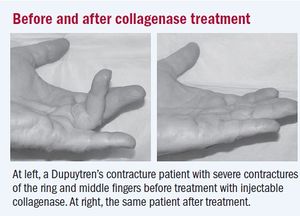
- Collagenase injections provide a minimally invasive treatment derived from Clostridium histolyticum. Night extension splinting is maintained for 6 months. Collagenase injections result in a 75% contracture reduction with a 35% recurrence charge per unit. Complications include edema, skin fierce, tendon rupture, complex regional pain syndrome, and pulley rupture. Earlier and later on collagenase treatment image at R [vii]
- Surgical fasciectomy can be either limited or radical. The recurrence rate at ane to ii years is xxx%, 15% at 3 to 5 years, and less than 10% subsequently ten years.
- Total palmar fasciectomy tin can besides be performed but is infrequently used as it requires resection of all palmar and digital fascia, including nondiseased tissue.
- Complications of fasciectomy include skin necrosis, hematoma (well-nigh common complexity), flare reaction, neurovascular injury, digital ischemia, swelling, and infection.
Irrespective of the treatment, recurrence is common with all of them, budgeted 20-50% at 5 years.[two]
Physical Therapy Direction [edit | edit source]
Conservative Approach [edit | edit source]
Concrete therapy may include ultrasound waves: oestrus (early stage of the disease); brace/splint to stretch the digits; a range of motion of the fingers to prevent adhesions.

Postoperative Intendance/Rehabilitation [edit | edit source]
Patients often enter hand therapy to :
- Maintain the range of move of the hand and fingers is important (for many activities of daily living), run across mitt exercises
- Extension splints often are used in conjunction with other modalities.
- Odema and scar interventions.[viii]
- Should be undertaken for at least three months to prevent contractures.
- Maximal benefits of surgery are not immediate, only get obvious later on half-dozen-8 weeks.[ii]
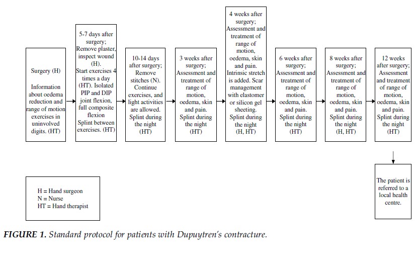
A standard protocol for postoperative management of Dupuytren disease is shown below (Engstrand et al. in 2009).[eight]
- Within the initial five days postoperative, the chief interventions are to educate the patient on decreasing edema and the importance of performing a range of motion exercises on the uninvolved fingers.
- Afterwards 5-7 days postoperative, the primary interventions shift to a range of movement exercises and splinting.
- The exercises are adapted to each subject area's individual goals and are based on their impairment, physical status, and competency.
- The types of splints used included volar splints, dynamic extension splint, dynamic flexion splints, exercise splints, and wrist splints.
The video below gives a expert summary of the condition and physiotherapy treatment (less than 4 minutes)
[9]
Conclusion [edit | edit source]
The cardinal fact to appreciate is that not all patients need handling.
- At that place are many treatments available for Dupuytren contracture and none is ideal or works consistently.
- But symptomatic patients should be offered treatment because all treatments take complications.
- The patient must be educated about the potential complications of treatments, which are worse than the disorder itself.
- Close advice between the team is essential in order to amend outcomes.
- Overall, only a few patients accomplish a desirable result.
- In many cases, prolonged physical therapy is required to restore functionality[ii]
References [edit | edit source]
- ↑ E Soreide, Thou H Murad, J M Denbeigh, East A Lewallen, A Dudakovic, L Nordsletten, A J van Wijnen, S Kakar.Handling of Dupuytren's contracture: a systematic review.PubMed.gov.National Library of Medicine. National Heart for Biotechnology Information.2018 Sep;100-B(9):1138-1145.doi: ten.1302/0301-620X.100B9.BJJ-2017-1194.R2.
- ↑ 2.0 2.1 ii.2 2.3 2.4 ii.5 2.half dozen 2.7 Walthall J, Rehman UH. Dupuytrens Contracture. InStatPearls [Internet] 2019 February nineteen. StatPearls Publishing.Bachelor from:https://www.ncbi.nlm.nih.gov/books/NBK526074/ (last accessed iv.four.2020)
- ↑ Bayat A. A nonsurgical therapy for Dupuytren illness. Rheumatology. 2010;6:vii-8.
- ↑ Dupuytren's Illness. American Society for Surgery of the Hand Web site. http://world wide web.assh.org/Public/HandConditions/Pages/DupuytrensDisease.aspx. 2010. Accessed March xix, 2011.
- ↑ 5.0 5.ane 5.two Walthall J, Anand P, Rehman UH. Dupuytren Contracture. StatPearls [Cyberspace]. 2020 Sep 14.
- ↑ Mayo Foundation for Education and Research. The Dupuytren's Contracture Folio. http://www.mayoclinic.com/wellness/dupuytrens-contracture/DS00732. Updated May 15, 2010. Accessed March 14, 2011.
- ↑ Harvard University. Nonsurgical approach unlocks contracted fingers. Harvard Women's Wellness Watch. 2009:6-vii.
- ↑ eight.0 eight.1 Engstrand C, Boren L, Liedberg GM. Evaluation of activity limitation and digital extension in Dupuytren's contracture 3 months after fasciectomy and paw therapy interventions. J Mitt Ther. 2009;22:21-27.
- ↑ Physio vibes DUPUYTREN'S CONTRACTURE & PHYSIOTHERAPY Direction Available from: https://www.youtube.com/lookout?v=a8KMCAFx8xw (final accessed 5.4.2020)
Source: https://www.physio-pedia.com/Dupuytren%E2%80%99s_Contracture
0 Response to "How Do You Know the Degree of a Dupuyten Contrature"
Post a Comment